Imagine holding a grain of sand between your fingertips. It is so small that you could lose it with a single careless movement, so ordinary that you would hardly give it a second thought. Yet inside a piece of mouse brain tissue no larger than that grain, scientists have found a universe. It contains hundreds of millions of connections, woven together in patterns so intricate that mapping them required years of work, enormous computing power, and a new kind of partnership between human scientists and artificial intelligence.
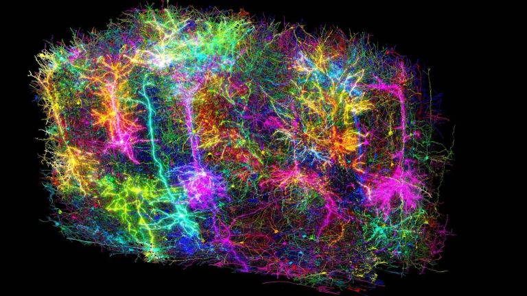
In April 2025, an international collaboration called MICrONS — Machine Intelligence from Cortical Networks — unveiled one of the most ambitious maps ever made of a living brain. The team reconstructed a small volume of mouse visual cortex in extraordinary detail, tracing neurons and their connections at a scale that had never before been attempted for a mammalian brain. The dataset contained roughly 523 million detected synapses, the tiny junctions where neurons pass signals to one another, and about 1.6 petabytes of data. To put that in perspective, 1.6 petabytes is more than a million gigabytes, all devoted to describing a speck of tissue smaller than a pea.
What makes this achievement remarkable is not only its size. For the first time at this scale, researchers linked what neurons do with how they are wired together. Before the tissue was examined under an electron microscope, the scientists recorded how many of those same neurons responded to visual scenes while the mouse was awake and watching. They could therefore ask a question that neuroscience has pursued for decades: does the wiring of the brain reflect what its cells actually compute? The early answer, emerging from the MICrONS data, is that it does — in ways that are both elegant and surprising.
This article explores what was discovered, how the map was made, and why it matters far beyond the laboratory. It is a story about vision, about the hidden architecture of thought, and about a new scientific resource that may change how we understand brains, build artificial intelligence, and study disease.
1. The Brain Is Not Simply a Collection of Neurons
A common picture of the brain shows isolated nerve cells with long, branching extensions, as if each cell worked alone. That picture is misleading. A neuron receives signals through tree-like structures called dendrites and sends signals along a projection called an axon. Where an axon meets another cell, a synapse forms. It is at those junctions — hundreds of millions of them in even a tiny volume — that information is transformed, amplified, filtered, and combined.
Consider what happens when you look at a moving bicycle. Light reflected from the bicycle enters your eyes and is converted into neural signals by the retina. Those signals travel to the visual cortex, a region at the back of the brain that processes what we see. Some neurons there respond strongly to edges of a particular orientation, others to movement in a particular direction, and still others to combinations of features that are harder to describe in words. No single neuron announces that a bicycle exists. Perception emerges from populations of cells, each contributing a partial piece of information, interacting through their connections.
This creates an extraordinary scientific challenge. If researchers record a neuron responding strongly to movement toward the right, they know something about its function, but not necessarily who it talks to. Conversely, a detailed picture of its physical connections does not automatically reveal what the neuron does. A complete explanation requires both structure and function, recorded in the same brain, at the same time, and linked cell by cell.
Think of a city. A map showing every street tells you where vehicles could travel, but not where people actually go, when traffic peaks, or which routes matter most. The brain presents a similar problem, except its roads are microscopic, its intersections can change their strength, and its traffic is the very stuff of perception. MICrONS was designed to connect those different kinds of knowledge, street map and traffic flow together.
2. What Scientists Actually Discovered
The MICrONS project focused on a small but important part of the mouse brain: several areas of visual cortex that work together to process what the animal sees. Researchers assembled a dataset containing roughly 523 million detected synapses, with the anatomical shapes of tens of thousands of neurons reconstructed in three dimensions. The amount of underlying information was staggering, not only because of its volume, but because it described different levels of complexity at once: the shape of each cell, the path of its axon, the location of its synapses, and, for a subset of cells, how it responded to visual input.
According to the United States National Institutes of Health, the project generated about 1.6 petabytes of data. Those numbers are difficult to visualize because they describe a tiny physical volume translated into an immense digital representation. Individual neurons had to be followed through thousands of thin slices of tissue, like tracing a single thread through a vast tapestry without losing track of where it goes, where it branches, and where it ends.
The truly distinctive achievement involved linking anatomy with activity. Before the tissue was reconstructed using electron microscopy, scientists recorded responses from neurons in the living mouse as it watched natural videos and other visual patterns. They could then match a subset of those functional recordings to cells identified in the anatomical reconstruction. That matching process offered an experimental bridge between questions that had traditionally been studied separately.
Which neurons respond to similar visual information? Are they more likely to form synapses with one another than with neurons that respond differently? These are not merely descriptive questions. They concern possible organizing principles of neural computation — the rules, if any, that shape how a brain wires itself to see the world.
A major companion study found evidence for a general pattern often described as like-to-like connectivity. Neurons with more similar visual responses were more likely to be connected than neurons with dissimilar responses. Importantly, this tendency appeared not only within individual cortical areas but also across layers and areas. That does not mean every similarly responding pair is connected, or that neural circuits follow one simple rule. It provides a foundation for testing why those patterns occur and what they accomplish.
3. Watching the Brain Before Mapping Its Wiring
The remarkable feature of MICrONS is that the researchers did not begin by examining only preserved tissue. They first observed neurons while the brain was functioning. An awake mouse was presented with visual stimuli, including natural videos and computer-generated patterns, while scientists recorded activity from thousands of neurons across multiple visual areas.
Neurons communicate electrically, but their activity can also be associated with changes in calcium concentration inside the cell. Scientists can introduce genetically encoded indicators that change their fluorescence in response to calcium, allowing activity to be watched with microscopes as glowing signals rise and fall. The resulting recordings do not represent direct measurements of every electrical impulse, and calcium signals are slower than the spikes they reflect. Nevertheless, they provide a powerful way to observe many neurons simultaneously, and to see which cells respond together to the same scene.
In the MICrONS experiments, researchers collected functional information across multiple visual areas. That detail matters because visual processing is not performed by one isolated region. Signals travel through interconnected cortical areas that represent and transform visual information in stages, from simple edges and motion to more complex patterns. By recording activity across several regions, the scientists could investigate how related responses were distributed and coordinated.
The next challenge was preserving the correspondence between those living neurons and their eventual anatomical reconstruction. Once the functional recordings were complete, the brain tissue could be prepared for electron microscopy. Tissue processing introduces deformation, and microscopy systems operate at dramatically different resolutions. Researchers therefore needed to align the datasets and identify which anatomical cells corresponded to which recorded cells. This is called co-registration, and it is one of the reasons the project required such careful, sustained work.
Imagine photographing a city from an airplane, then dismantling the entire city into microscopic components and trying to match each building in the photograph to its pieces. The comparison is imperfect, but it captures the fundamental difficulty. The project did not establish functional recordings for every neuron in the anatomical dataset, nor did it claim to. Instead, it created a large and carefully documented overlap between two different kinds of scientific evidence, and that overlap became the foundation for examining the relationship between structure and vision.
4. How Researchers Turned Microscopic Tissue Into a Digital Map
After the living-brain recordings, scientists faced another enormous obstacle: reconstructing the physical wiring. Ordinary photography cannot accomplish this. Synaptic structures are far smaller than anything visible to the naked eye, and many of the relevant branches are thinner than a wavelength of light. Researchers therefore relied on electron microscopy, which uses electrons rather than visible light to image tissue at nanometer resolution.
The process involved cutting the brain tissue into thousands of extraordinarily thin sections and imaging each one in detail. Each individual image captured only part of the biological structure. A neuron's axon might appear in one section, continue into another, branch across multiple images, and connect to many different partners along the way. To reconstruct the full pathway, software had to follow those structures through the stack of images, section after section, without confusing one branch for another.
This is where artificial intelligence became essential. Machine-learning algorithms helped identify cellular boundaries, segment neural structures, detect connections, and follow branches through the image stack. Without automation, tracing the immense network manually would have been impractical, taking far longer than any research team could sustain. However, AI did not eliminate the need for human expertise. Microscopic images contain ambiguities. Two branches can pass close together without connecting. Boundaries can be faint, and a single error can create a false connection or miss a real one.
Such errors matter because a false connection can alter conclusions about circuit organization. Researchers therefore used combinations of automated correction and manual proofreading, paying particular attention to cells and connections that were central to their analyses. The process demonstrates an important principle about AI-assisted discovery. Machine learning can accelerate the extraction of patterns from enormous datasets, but scientific conclusions still depend on careful validation, transparent methods, and an understanding of where errors are most likely.
The finished connectome is best understood as a large, evolving scientific reconstruction rather than a perfect, final photograph. Nevertheless, its scale transformed what researchers could investigate. Instead of reconstructing a handful of selected cells, scientists gained access to a dense anatomical context. They could examine not only whether particular cells were connected, but also how those connections compared with the connections that were possible but absent. That comparison is crucial, because without knowing which connections were possible, researchers risk mistaking patterns caused by simple proximity for patterns caused by functional organization.
5. The Hidden Rule: Similar Neurons Often Connect
One of the most intriguing scientific findings concerns a simple question: do neurons connect randomly, or do they follow rules? The answer is not a straightforward choice between complete randomness and perfect organization. Brain circuits contain both structured patterns and substantial variability, and MICrONS helps quantify that balance.
In the mouse visual cortex, researchers found that neurons with more similar visual response properties were more likely to be connected. This relationship is often described as like-to-like connectivity. Imagine a large group of musicians performing in an orchestra. Some musicians specialize in similar instruments or musical roles. Although everyone contributes to the whole performance, those with similar roles often coordinate closely with one another. In a visual circuit, neurons that respond similarly to visual stimulation may also participate in related computations, and connections between those cells can help coordinate or transform signals.
But the discovery becomes more interesting when the brain's anatomy is considered. Neurons are not arranged in a flat sheet where every cell has equal access to every other cell. They occupy different cortical layers and regions. Their branches extend in complex directions, and physical proximity alone does not guarantee a connection. A neuron might be functionally similar to another neuron but physically unable to form a direct connection, or it might be near many cells it never connects to.
Researchers therefore compared connected neurons with nearby potential partners that were not connected. By analyzing connectivity alongside functional similarity, they could examine whether like-to-like relationships held up after accounting for distance and anatomy. The results supported a broader organizing tendency across local and inter-area pathways. That finding does not imply that a visual cortex is neatly divided into isolated groups representing single features, nor does it mean that every neuron receiving similar visual information performs an identical task. Neural computation relies on excitation, inhibition, recurrent interactions, and many forms of modulation. The significant point is that circuit organization appears statistically related to what neurons respond to, and that gives scientists something more useful than a beautiful map: a hypothesis that can be tested.
Future experiments can examine whether these connections arise during development, change with visual experience, or differ between brain areas. The discovery also invites comparisons with artificial neural networks, where patterns of connectivity are learned rather than grown, and where architecture is designed rather than evolved.
6. A Digital Twin of Visual Activity
Another important development emerged from the project's use of predictive artificial intelligence. Researchers built computational models designed to predict how neurons in visual cortex would respond to new visual inputs they had never seen. These models were trained using large datasets of activity collected while mice watched videos and other patterns. The objective was not to reproduce the anatomy of the brain perfectly. Instead, the models attempted to learn relationships between visual inputs and neuronal responses.
Consider a simple example. Suppose a particular neuron responds strongly when a vertical edge moves across part of the visual field. A computational model might learn to predict that response from images containing similar features. But real neurons often respond to combinations of features, contexts, and movements that are difficult to describe with simple rules. Modern deep-learning systems can learn complicated relationships directly from large collections of examples, and in the MICrONS-related research, a foundation model of visual cortical activity was evaluated on its ability to generalize to new stimuli, including different forms of visual patterns and moving scenes.
The results showed meaningful generalization, suggesting that models trained on sufficiently rich neural data can predict responses to stimuli they were not trained on. Researchers also used this modeling framework to investigate relationships between predicted neural activity and anatomical structure. That is where the term digital twin enters the discussion. In engineering, a digital twin usually refers to a computational representation of a physical system that can be used to simulate its behavior. In this case, the digital representation concerns neuronal responses and relationships derived from real data.
It is not a complete functioning copy of a mouse brain. The model does not recreate the animal's memories, motivations, consciousness, or full nervous system. It also does not demonstrate that the brain's biological machinery can be replaced by a conventional computer. Its purpose is narrower and scientifically valuable. The model offers researchers a way to generate predictions about how neural populations respond under new conditions. Those predictions can then be compared with experimental recordings. A successful prediction provides evidence that the model captures something useful about the system. A failed prediction is also useful, because it shows where the model is wrong and where new experiments are needed.
This approach is especially promising when combined with the connectome. Researchers can ask whether a cell's physical position, shape, connectivity, or relationship to other cells helps predict its function. Over time, such experiments may help distinguish features of neural architecture that are essential for vision from features that are incidental.

7. Why This Discovery Matters for Artificial Intelligence
Artificial intelligence has made remarkable progress by learning patterns from large datasets, but real brains still do many things that artificial systems struggle with. Modern AI systems can recognize images, translate languages, generate text, and solve complex problems, yet they often require enormous amounts of data and energy, and they can fail in ways that seem surprisingly brittle compared with animal perception. The MICrONS dataset offers an opportunity to study neural computation from the biological side of that comparison.
One important question concerns connectivity. Many artificial neural networks use mathematical operations that connect computational units according to patterns learned from data. Biological networks, by contrast, develop through interactions involving genes, activity, experience, and the physical constraints of growing tissue. Understanding those constraints could reveal principles that artificial systems have not yet exploited. For example, biological circuits often process information through recurrent connections, where activity loops back and influences later processing. Researchers can investigate whether specific recurrent wiring patterns support particular visual computations, such as tracking motion or recognizing an object despite changes in lighting.
Another question involves efficiency. The brain performs demanding sensory computations while operating within biological limits on energy and space. That does not automatically mean every biological circuit is more efficient than every artificial system, and comparisons must be made carefully. Nevertheless, discovering how neural circuits balance connectivity, information processing, and metabolic cost could inspire more efficient designs. There is also the challenge of robustness. Animals can recognize familiar objects under changing lighting, motion, distance, and partial obstruction, often from just a few examples. Detailed maps can help researchers ask whether certain arrangements of neurons contribute to stable perception.
The most interesting outcome may not be a direct copy of brain architecture. It could be the discovery of abstract principles that engineers can adapt without reproducing the biology literally. An airplane does not flap its wings like a bird, yet studying flight in nature helped inspire fundamental ideas about lift and control. Similarly, understanding neural circuitry might suggest computational strategies rather than literal blueprints. At the same time, comparisons must remain disciplined. A correlation between neuronal connectivity and visual responses does not establish that an analogous artificial architecture will work better. Such claims require experiments in which proposed principles are implemented, tested against alternatives, and evaluated on tasks that matter.
8. Could Brain Maps Help Us Understand Disease?
Many neurological and psychiatric conditions involve changes in brain function, communication, development, or connectivity. It is therefore reasonable to ask whether more detailed maps of neural circuits could contribute to understanding them. The answer is potentially yes, but the path from foundational discoveries to treatments is neither short nor straightforward.
Consider epilepsy. Seizures involve abnormal patterns of neuronal activity that can spread through brain networks. Detailed circuit maps could eventually improve the models scientists use to study how particular patterns emerge and spread. However, a mouse visual-cortex connectome is not a direct model of every form of human epilepsy. The relevant circuits, diseases, developmental histories, and clinical circumstances can differ substantially. A similar distinction applies to Alzheimer's disease. Researchers know that Alzheimer's involves complex pathological changes, including alterations in synaptic function, but a reference map is not a diagnostic test or treatment.
For conditions involving neurodevelopment, such as autism, circuit-level research may help scientists understand how differences in connectivity relate to differences in perception and behavior. It would be scientifically inappropriate, however, to treat such conditions as consequences of one simple wiring error. Brains exhibit enormous variation, and clinical conditions involve many interacting biological and environmental factors. The potential contribution of connectomics is therefore more fundamental. It may allow scientists to construct more realistic hypotheses about how cellular changes influence network behavior, and those predictions could guide experiments and, in the long term, inform therapeutic strategies. The strongest medical applications will likely emerge through combinations of connectomics, genetics, physiology, and clinical research. A wiring diagram alone cannot explain a disease, but without understanding the healthy architecture of neural communication, some disease mechanisms are harder to see.
9. The Limits of a Map: Why Synapses Are Not the Whole Story
The enormous scale of the MICrONS dataset can create a misleading impression that researchers are approaching a complete explanation of the brain. The reality is more complicated. A synapse is not simply a fixed wire. Its effectiveness can change depending on recent activity, molecular conditions, receptor availability, and the state of the surrounding tissue. Some synapses strengthen or weaken through mechanisms associated with learning and experience. Others are influenced by chemical signals and changing physiological states. Two circuits with similar anatomical arrangements may therefore operate differently because their synapses behave differently.
A static anatomical reconstruction captures physical relationships at a particular moment. It does not automatically reveal the precise strength of every synapse, the timing of every electrical signal, or the influence of modulatory chemicals that can reshape network activity. The brain also contains important non-neuronal cells. Astrocytes, microglia, oligodendrocytes, and other cell populations contribute to brain development, maintenance, and signaling in ways that a neuron-only map cannot fully capture. Understanding neural computation requires more than tracing axons and dendrites.
There are additional limits involving scale. The MICrONS project studied a portion of the mouse visual cortex. It did not reconstruct the entire mouse brain, let alone a human brain. Human brains are much larger and contain diverse circuits involved in language, memory, movement, emotion, and planning. Even if every synapse in a human brain could eventually be mapped, scientists would still need to explain how activity across those circuits gives rise to experience. A complete anatomical map would be a monumental achievement, but it would not automatically explain consciousness, which remains an active field of scientific and philosophical investigation. It is important not to confuse the ability to reconstruct neural connectivity with the ability to recreate a mind.
There are also practical limitations within the dataset itself. Some reconstructions require proofreading, certain synapses may be missed or incorrectly detected, and functional recordings cover only a subset of cells. These limitations do not diminish the achievement. They define the conditions under which its results should be interpreted, and they point to where future work is needed. Scientific progress often depends on understanding precisely what a measurement can and cannot establish, and in this case the most responsible interpretation is also the most exciting: researchers have gained an unprecedented, shared window into brain organization.
10. An Open Scientific Resource That Could Accelerate Discovery
One of the most important aspects of MICrONS is that its researchers made extensive datasets and analytical tools publicly available. This changes who can investigate the questions raised by the project. Traditionally, advanced electron microscopy and large-scale neural reconstruction have required extraordinary resources and specialized facilities. A publicly accessible dataset does not eliminate those barriers entirely, but it can reduce the need for every team to repeat the most expensive steps.
A researcher studying visual processing can investigate connections among selected cell types without building a new microscope facility. A computational neuroscientist can develop algorithms to identify anatomical patterns and compare them with functional data. A machine-learning researcher can investigate whether neural morphology contains information about function that models can learn. Students can learn how real brain datasets are organized and how uncertainty affects scientific conclusions. Researchers from different institutions can also test competing hypotheses using shared evidence, which is especially important for a field as complex as neuroscience.
If different teams study different animals, brain regions, techniques, and conditions, their findings can be difficult to compare. Shared datasets make it possible to examine multiple explanations within the same experimental framework. For example, one team might argue that connectivity is primarily explained by physical proximity, while another argues that functional similarity matters more. Both approaches can be evaluated against the same anatomical and functional measurements, and the results can then be challenged, extended, or refined by other researchers. This does not guarantee consensus, but it creates better opportunities for identifying where scientific disagreements actually lie.
Open scientific resources also encourage the development of improved analytical tools. A reconstruction algorithm that performs well on one carefully selected example may fail when confronted with the full complexity of a large dataset. Large datasets expose those weaknesses, and software developers can test new segmentation, visualization, proofreading, and network-analysis methods against real, demanding data. The wider significance is that MICrONS is not simply a collection of published conclusions. It is an experimental foundation upon which additional discoveries can be built, and its scientific value may continue to grow as computational techniques improve and researchers ask questions the original team never considered.
11. What Happens Next: From Wiring Diagrams to Explanations
The next generation of connectomics research will face a challenge that is more difficult than producing ever-larger maps. Scientists must determine which aspects of those maps explain how neural circuits behave. One promising approach involves combining anatomical reconstructions with targeted experiments. Imagine a model predicting that a particular group of connected neurons is important for distinguishing one kind of motion from another. Researchers could test that prediction by selectively altering activity in the circuit and measuring whether perception changes in the predicted way. If the predicted changes occur, the evidence for the model becomes stronger. If they do not, researchers must reconsider the proposed mechanism. This movement from description to prediction and experimental testing is essential.
Another direction involves comparing circuits across different animals and across development. The MICrONS dataset includes information from one mouse's reconstructed visual-cortex volume. Repeated measurements could reveal whether like-to-like connectivity is a broadly conserved organizing principle, or whether it varies with species, age, experience, or brain area. Development is especially interesting, because the adult brain contains networks shaped by many influences, including spontaneous activity, sensory experience, and learning. A static reconstruction cannot explain exactly how those connections formed, but comparisons across developmental stages could help researchers investigate the processes that produce them.
Another challenge is bridging spatial scales. A synapse operates at a microscopic level, while perception involves distributed activity across large networks. Scientists need models capable of connecting those levels without becoming computationally impossible or biologically meaningless. Future work may combine structural reconstructions, electrophysiological recordings, optical imaging, and computational modeling, with artificial intelligence remaining an important tool for identifying patterns in data too large for manual analysis. Yet the ultimate aim should not be simply to accumulate more data. The goal is to discover explanations that make accurate predictions about real biological systems — explanations that tell researchers why a particular network behaves as it does, what would happen if it were changed, and how it might fail.
Conclusion: The Map Is Only the Beginning
For centuries, humans have looked toward distant galaxies, deep oceans, and unfamiliar landscapes in search of new frontiers. Yet one of the most complicated frontiers has always been inside our own heads. The MICrONS project reveals just how intricate that frontier is. Within a microscopic portion of mouse brain tissue, scientists reconstructed a network containing hundreds of millions of synapses, linked — for a subset of cells — to the visual responses those cells produced while the animal was alive.
The achievement required experimental innovation, artificial intelligence, extraordinary computing resources, and years of collaboration across institutions. Its central lesson is that understanding the brain requires more than identifying individual cells. We must investigate how those cells are connected, how they influence one another, and how their collective activity gives rise to perception. The findings suggest that neural wiring is not arbitrary. Relationships between connectivity and function appear to follow statistical patterns that can be tested, refined, and perhaps one day explained.
At the same time, the project reveals how much remains unknown. The brain is not a fixed electronic circuit. It is a living system whose connections, signals, chemistry, and cellular partners change over time. A detailed map brings scientists closer to understanding that complexity, but it does not make the complexity disappear. Perhaps the most remarkable aspect of this discovery is the contrast between its physical scale and its scientific ambition. A volume comparable to a grain of sand required a digital dataset of staggering size, and even that dataset represents only a small part of one brain.
The future of neuroscience may depend on learning how to move between these two perspectives: the intimate, microscopic detail of a single synapse, and the broad, distributed activity that lets an animal see, learn, and act. The map has opened a door. What remains is to discover how the machinery behind it turns light into perception, structure into function, and wiring into understanding — and that work, like the map itself, is only beginning.
Sources
- MICrONS Consortium (2025). Functional connectomics spanning multiple areas of mouse visual cortex. Nature.
- National Institutes of Health (April 9, 2025). Scientists map unprecedented detail of connections in visual perception in the mouse brain. NIH News Releases.
- MICrONS-related research (2025). Functional connectomics reveals general wiring rule in mouse visual cortex. Nature.
- MICrONS-related research (2025). Foundation model of neural activity predicts response to new stimuli. Nature.
- Nature (April 9, 2025). The MICrONS Project: an explanation of the collaboration, dataset, and scientific goals.
- MICrONS Explorer. Public cortical connectomics datasets, analytical tools, documentation, and visualizations.
- National Institutes of Health (2024). Unseen details of human brain structure revealed. Background research.
The conversation
Post a Comment